

Self-delivering AUMsilence sdASO
Targeted mRNA knockdown, from cell culture to in vivo studies
No lipid-based transfection reagent, electroporation, or viral vector required.
In an animal the same AUMsilence
T cells
CD4+ (naive, activated, memory, exhausted), CD8+ (naive, effector, memory, exhausted), Tregs, Th1, Th2, Th17, Tfh, gd T cells, NKT, MAIT, CAR-T cells, TILs
B cells and NK cells
Naive B cells, Memory B cells, Plasma cells, GC B cells, Primary NK cells (CD56+CD3-), CAR-NK cells
Macrophages, DCs and granulocytes
Monocytes, M1/M2 macrophages, BMDMs, Peritoneal, Alveolar, Kupffer cells, Microglia, TAMs, Myeloid DCs, Plasmacytoid DCs, Neutrophils, Eosinophils, Basophils, Mast cells
Hematopoietic progenitors
Long-term HSCs, Short-term HSCs, CD34+ HSPCs, MPPs, CLPs, CMPs, GMPs, MEPs, AGM embryonic HSCs
Neurons
Cortical, Hippocampal, Motor, Sensory, DRG, Dopaminergic, GABAergic, Glutamatergic, Purkinje, Retinal ganglion, Enteric, iPSC-derived neurons
Glial and neural stem cells
Astrocytes, Oligodendrocytes, Microglia, Schwann cells, hiPSC-derived NSCs, Adult neural progenitors, Hippocampal slices, Brain sections, Spinal cord explants
Cardiovascular
Primary and iPSC cardiomyocytes, HASMCs, HCASMCs, Pulmonary artery SMCs, Endothelial cells (HUVECs, HAECs), Cardiac fibroblasts, Pericytes
Musculoskeletal
Myoblasts, Myotubes, Satellite cells, Osteoblasts, Osteoclasts, Osteocytes, Chondrocytes, Synoviocytes, Tenocytes, MSCs
Stem and progenitor cells
Human ESCs, Mouse ESCs, iPSCs, iPSC-derived neurons, cardiomyocytes, hepatocytes, NSCs, HSCs, CD34+ HSPCs, MSCs, Cardiac and Endothelial progenitors, Intestinal stem cells (Lgr5+)
Liver and gastrointestinal
Primary hepatocytes, Hepatic stellate cells, Cholangiocytes, Intestinal epithelial, Colonocytes, Paneth cells, Goblet cells, Pancreatic beta, ductal, and acinar cells
Lung, airway and kidney
Tracheobronchial epithelial, Alveolar epithelial type I and II, Bronchial epithelial, Airway smooth muscle, Lung fibroblasts, Podocytes, Mesangial cells, Tubular epithelial cells
Skin, eye, reproductive and other
Keratinocytes, Melanocytes, Fibroblasts, CAFs, RPE cells, Corneal epithelial, Trophoblasts, HEK293T, NIH3T3, THP-1, Jurkat, Neuro-2a
- No transfection reagents
- No viral vectors
- No electroporation
- Suspension and adherent cells
Tumor models
Tumor spheroids (any cancer type), Patient-derived tumoroids (CRC, lung, liver, pancreatic, breast, ovarian), PDX-derived organoid cultures, Multicellular tumor microenvironment models, Co-culture systems (immune cells + tumor cells)
CNS organoids
Brain organoids, Cerebral organoids, Spinal cord organoids, Retinal organoids, Cortical organoids, Midbrain organoids
Organ organoids
Intestinal organoids, Kidney organoids, Liver and hepatic organoids, Lung and airway organoids, Cardiac organoids, Gastric organoids, Pancreatic organoids, Prostate organoids
Tissue-engineered models
3D microvasculature (HUVECs on beads), Air-liquid interface (ALI) cultures, Organ-on-chip models, Tissue explant cultures (brain, muscle, AGM), Transwell co-culture and migration systems, Scaffold-based 3D cultures
Solid tumors
Melanoma, Glioblastoma (GBM), DIPG/DMG, Prostate cancer, Breast cancer, Ovarian cancer, NSCLC, SCLC, Hepatocellular carcinoma (HCC), Cholangiocarcinoma, Colorectal cancer, Gastric cancer, Esophageal cancer, Renal cell carcinoma, Bladder cancer, Pancreatic cancer, Head and neck SCC, Thyroid cancer, Sarcomas, Mesothelioma
Hematologic malignancies
Acute lymphoblastic leukemia (B-ALL, T-ALL), Acute myeloid leukemia (AML), Chronic myeloid leukemia (CML), Chronic lymphocytic leukemia (CLL), Diffuse large B-cell lymphoma (DLBCL), Burkitt lymphoma, Hodgkin lymphoma, Mantle cell lymphoma, T-cell lymphoma, Multiple myeloma, Thymoma
Patient-derived models
Patient-derived neurospheres (GBM, DMG), Patient-derived tumoroids (CRC, lung, liver, breast), Tumor-infiltrating lymphocytes (TILs) from surgical specimens, Primary cancer cells from patient biopsies, PDX-derived cell cultures
- AUMsilence sdASOs penetrate 3D structures without carriers
- Compatible with organoid protocols
In vivo administration routes
Optimize administration route, formulation, dose, administration volume, and dosing schedule for each study.
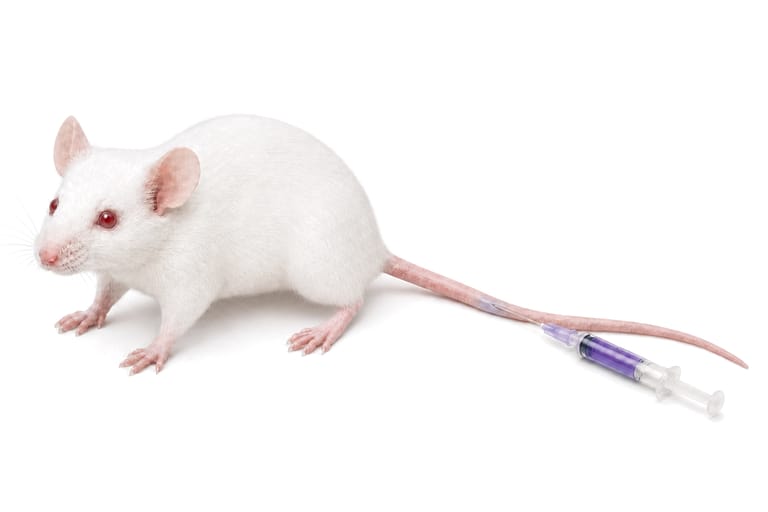
Route 1
Intravenous IV
Lateral tail vein
Lateral tail vein: a fine needle nearly parallel to the tail, entering the distal third first.
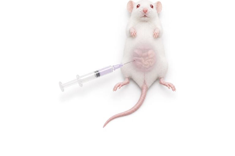
Route 2
Intraperitoneal IP
Right lower abdominal quadrant
Lower right quadrant of the abdomen, the needle toward the head at 30 to 45 degrees, at most 10 mL/kg.
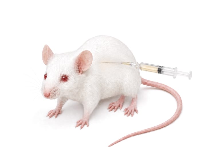
Route 3
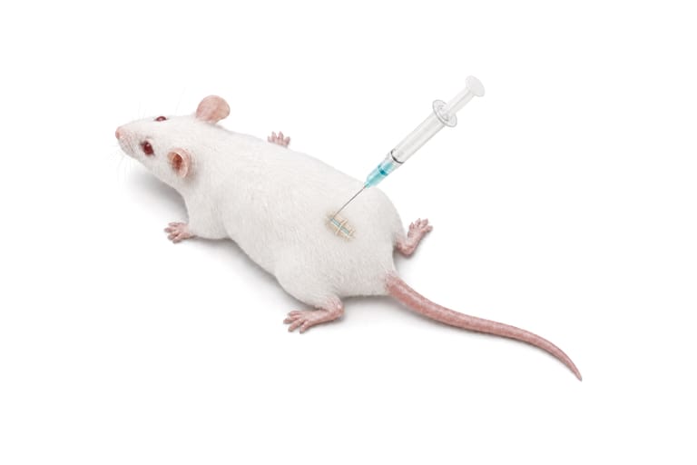
Subcutaneous SC
Dorsolateral flank
Dorsolateral flank: the skin tented, the needle slid under the skin parallel to the back.
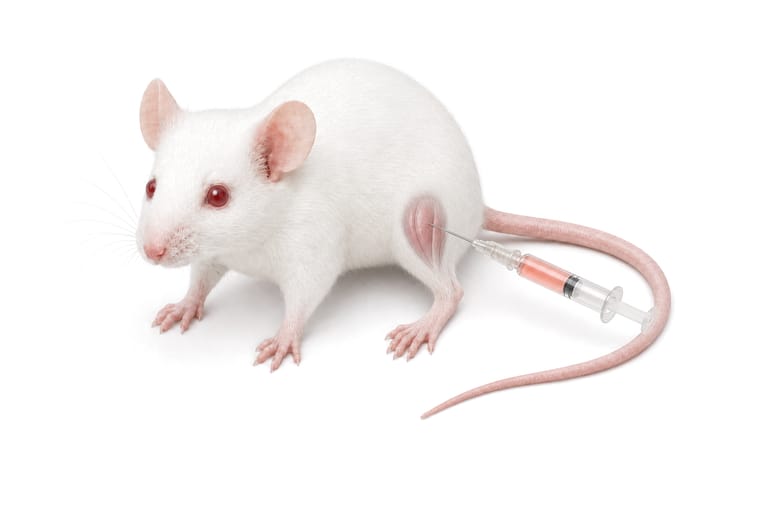
Route 4
Intramuscular IM
Proximal quadriceps muscle
Front of the thigh, the quadriceps, clear of the sciatic nerve; a fine needle, tens of microliters.
Route 5
Intrathecal injection IT
Lower lumbar L5-L6 interspace
Lumbar puncture between L5 and L6, a 30-gauge needle, 10 microliters, the tail flick as the sign.
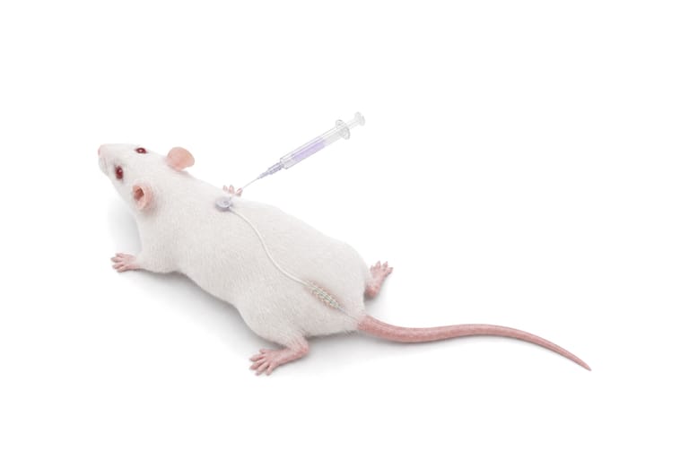
Route 6
Intrathecal catheter IT
Lumbar catheter and dorsal port
A lumbar catheter placed between L5 and L6 and advanced to the lumbar cord, dosed through a dorsal port.


For research use only. Not for use in diagnostic or therapeutic procedures.
Sources
- The pictures are the object pictures of AUM BioTech's workflow slide, rendered views of the objects on a white ground; none is a photograph of an experiment and none records a measurement.
- The pictures are rendered illustrations of each procedure, not photographs; the captions describe the procedure.
- Every number in a route's caption, the angle and the volume, the gauge and the vertebral level, is stated by the papers listed under Sources, each with the sentence it supports. The site named under each route, and the anatomy and the hand's technique a route's caption describes, are AUM BioTech's, as its workflow slide states them; the papers listed for such a caption carry the route and its numbers, not every clause, and the one paper that describes a catheter dosed through a port describes it in dogs.
- [R01] Galal S, Chaltel Lima L, Wang N, Moury C, Yan G, Dai M, et al. (2026). In vivo CRISPR screening identifies metastasis suppressors in triple-negative breast cancer. Nature Communications 17:9371. PMID 42680732, doi 10.1038/s41467-026-76293-x.Cited for Route 01, Intravenous: Lateral tail vein: a fine needle nearly parallel to the tail, entering the distal third first.
- [R02] Breuer CB, Xiong Z, Wang A, Rodriguez GE, Abhiraman GC, Garcia KC, et al. (2025). Spontaneous and experimental models of lymph node metastasis. Nature Protocols 20:3170-3187. PMID 40804176, doi 10.1038/s41596-025-01200-5.Cited for Route 01, Intravenous: Lateral tail vein: a fine needle nearly parallel to the tail, entering the distal third first.Cited for Route 03, Subcutaneous: Dorsolateral flank: the skin tented, the needle slid under the skin parallel to the back.
- [R03] Al Shoyaib A, Archie SR, Karamyan VT (2019). Intraperitoneal Route of Drug Administration: Should it Be Used in Experimental Animal Studies?. Pharmaceutical Research 37:12. PMID 31873819, doi 10.1007/s11095-019-2745-x.Cited for Route 02, Intraperitoneal: Lower right quadrant of the abdomen, the needle toward the head at 30 to 45 degrees, at most 10 mL/kg.
- [R04] Rysanek D, Chrienova Z, Stary D, Bajda M, Novak J, Vasicova P, et al. (2026). In silico screening, synthesis, and biological evaluation of pyrazolopyrimidine-derived mTOR inhibitors for anticancer and senomorphic effects. Cancer Cell International 26:247. PMID 42092963, doi 10.1186/s12935-026-04308-0.Cited for Route 02, Intraperitoneal: Lower right quadrant of the abdomen, the needle toward the head at 30 to 45 degrees, at most 10 mL/kg.
- [R05] Sagarbarria I, Seo J, Smith AJ, Vazey EM, Tremblay KD, Mager J, et al. (2026). Optimized detection of caspase-6 activation in a murine inflammation model to inform neurodegenerative disease therapies. PLOS ONE 21:e0351312. PMID 42308231, doi 10.1371/journal.pone.0351312.Cited for Route 02, Intraperitoneal: Lower right quadrant of the abdomen, the needle toward the head at 30 to 45 degrees, at most 10 mL/kg.
- [R06] Zhang P, Zhuang J, Hao Y, Zhu X, Liang H, Hu G, et al. (2026). GNPs-pIL-4 reprograms macrophage polarization and activates the OSM/GSNOR/ENG axis to improve angiogenesis in ischemic limbs. Science Advances 12:eaed5692. PMID 42585315, doi 10.1126/sciadv.aed5692.Cited for Route 02, Intraperitoneal: Lower right quadrant of the abdomen, the needle toward the head at 30 to 45 degrees, at most 10 mL/kg.
- [R07] E-Y Chuang A, Tung SY, Tzou KY, Cai YE, Hsu HC, Tsui KH, et al. (2026). A CD44-targeted PDA/ZnO redox-active nanocomposite that couples chemotherapy with mitochondrial stress and tumor immune remodeling. Journal of Nanobiotechnology 24:840. PMID 42702711, doi 10.1186/s12951-026-04899-w.Cited for Route 03, Subcutaneous: Dorsolateral flank: the skin tented, the needle slid under the skin parallel to the back.
- [R08] Wohlgemuth RP, Sriram S, Brashear SE, Henricson KE, Howard JJ, Smith LR (2026). Targeting muscle fibrosis using non-specific collagenase injections destabilizes the basal lamina and induces matrix remodeling. Skeletal Muscle 16:33. PMID 42321868, doi 10.1186/s13395-026-00432-7.Cited for Route 04, Intramuscular: Front of the thigh, the quadriceps, clear of the sciatic nerve; a fine needle, tens of microliters.
- [R09] Dos Santos M, Bezprozvannaya S, McAnally JR, Cai C, Liu N, Olson EN (2025). A mechanistic basis of fast myofiber vulnerability to neuromuscular diseases. Cell Reports 44:115959. PMID 40632651, doi 10.1016/j.celrep.2025.115959.Cited for Route 04, Intramuscular: Front of the thigh, the quadriceps, clear of the sciatic nerve; a fine needle, tens of microliters.
- [R10] Lee DE, McKay LK, Bareja A, Li Y, Khodabukus A, Bursac N, et al. (2022). Meteorin-like is an injectable peptide that can enhance regeneration in aged muscle through immune-driven fibro/adipogenic progenitor signaling. Nature Communications 13:7613. PMID 36494364, doi 10.1038/s41467-022-35390-3.Cited for Route 04, Intramuscular: Front of the thigh, the quadriceps, clear of the sciatic nerve; a fine needle, tens of microliters.
- [R11] Hylden JL, Wilcox GL (1980). Intrathecal morphine in mice: a new technique. European Journal of Pharmacology 67:313-316. PMID 6893963, doi 10.1016/0014-2999(80)90515-4.Cited for Route 05, Intrathecal injection: Lumbar puncture between L5 and L6, a 30-gauge needle, 10 microliters, the tail flick as the sign.
- [R12] Guo R, Chen O, Zhou Y, Bang S, Chandra S, Li Y, et al. (2025). Arrestin-biased allosteric modulator of neurotensin receptor 1 alleviates acute and chronic pain. Cell 188:4332-4349.e21. PMID 40393456, doi 10.1016/j.cell.2025.04.038.Cited for Route 05, Intrathecal injection: Lumbar puncture between L5 and L6, a 30-gauge needle, 10 microliters, the tail flick as the sign.
- [R13] Xu J, Yan Z, Bang S, Velmeshev D, Ji RR (2025). GPR37L1 identifies spinal cord astrocytes and protects neuropathic pain after nerve injury. Neuron 113:1206-1222.e6. PMID 39952243, doi 10.1016/j.neuron.2025.01.012.Cited for Route 05, Intrathecal injection: Lumbar puncture between L5 and L6, a 30-gauge needle, 10 microliters, the tail flick as the sign.
- [R14] Xu J, Li Y, Novak C, Lee M, Yan Z, Bang S, et al. (2026). Mitochondrial transfer from glia to neurons protects against peripheral neuropathy. Nature 650:951-960. PMID 41501451, doi 10.1038/s41586-025-09896-x.Cited for Route 05, Intrathecal injection: Lumbar puncture between L5 and L6, a 30-gauge needle, 10 microliters, the tail flick as the sign.
- [R15] Qian Y, Wang Y, Liu Y, Geng Y, Dong M, Huang Y, et al. (2026). Integrated analysis of single-cell and transcriptome data reveals temporal dynamics of microglial phagocytic reprogramming in neuropathic pain progression. Scientific Reports 16:27944. PMID 42706380, doi 10.1038/s41598-026-69062-9.Cited for Route 05, Intrathecal injection: Lumbar puncture between L5 and L6, a 30-gauge needle, 10 microliters, the tail flick as the sign.
- [R16] Wu WP, Xu XJ, Hao JX (2004). Chronic lumbar catheterization of the spinal subarachnoid space in mice. Journal of Neuroscience Methods 133:65-69. PMID 14757346, doi 10.1016/j.jneumeth.2003.09.015.Cited for Route 06, Intrathecal catheter: A lumbar catheter placed between L5 and L6 and advanced to the lumbar cord, dosed through a dorsal port.
- [R17] Luo X, Wickman JR, DaCunza JT, Tian Y, Sacan A, Ajit SK (2026). Microglial Epigenetic Memory is Associated with Accelerated Resolution of Inflammatory Pain Induced by Prophylactic Macrophage-Derived Small Extracellular Vesicles. Journal of Inflammation Research 19:598095. PMID 42445356, doi 10.2147/jir.s598095.Cited for Route 06, Intrathecal catheter: A lumbar catheter placed between L5 and L6 and advanced to the lumbar cord, dosed through a dorsal port.
- [R18] Blizzard CA, Lee KM, Dickson TC (2016). Inducing Chronic Excitotoxicity in the Mouse Spinal Cord to Investigate Lower Motor Neuron Degeneration. Frontiers in Neuroscience 10:76. PMID 26973454, doi 10.3389/fnins.2016.00076.Cited for Route 06, Intrathecal catheter: A lumbar catheter placed between L5 and L6 and advanced to the lumbar cord, dosed through a dorsal port.
- [R19] Boyd MM, Mazur C, Pribnow K, Zanon RL, Adams E, DuGal J, et al. (2025). Comparison of MALAT1 antisense oligonucleotide distribution following intracerebroventricular and lumbar intrathecal routes of administration. Molecular Therapy Nucleic Acids 36:102742. PMID 41216427, doi 10.1016/j.omtn.2025.102742.Cited for Route 06, Intrathecal catheter: A lumbar catheter placed between L5 and L6 and advanced to the lumbar cord, dosed through a dorsal port.
In vivo delivery, no carrier required
| Delivery route | Description | Research models and targets |
|---|---|---|
| Intraperitoneal (IP) | Systemic peritoneal delivery | Liver, spleen, immune cells, tumor models |
| Intravenous (IV) | Systemic vascular delivery | Liver, spleen, circulating cells, metastasis models |
| Intrathecal | Spinal canal / CSF delivery | Spinal cord injury, ALS, SMA, pain models |
| Stereotaxic (brain) | Direct CNS parenchymal injection | Alzheimer's, Parkinson's, tauopathies, brain tumors |
| Intracerebroventricular | CSF / ventricular delivery | CNS-wide distribution, neurodegenerative diseases |
| Intratumoral | Direct tumor injection | Solid tumors, xenograft models, syngeneic tumors |
| Subcutaneous | Local tissue delivery | Tumor models, fibrosis, local inflammation |
| Intranasal | Non-invasive CNS access | Brain delivery via olfactory route, respiratory targets |
| Intravitreal | Intraocular delivery | Retinal degeneration, macular degeneration, glaucoma |
| Intra-articular | Joint cavity delivery | Osteoarthritis, rheumatoid arthritis, cartilage repair |
| Topical / transdermal | Skin surface delivery | Dermatologic conditions, wound healing, psoriasis |
| Ex vivo to transplant | Cell treatment then infusion | HSC transplant, adoptive T cell therapy, CAR-T/NK |
- No delivery vehicle
- No lipid nanoparticles
- No viral vectors
- Direct administration in saline or PBS
Protocol
In vitro workflow
Optimize cell density, AUMsilence sdASO concentration, exposure duration, and assay time point for each model.
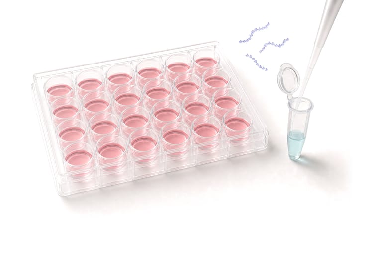
Step 1
Prepare cells and AUMsilence sdASO
Plate cells at the optimized density. Reconstitute AUMsilence sdASO in sterile, nuclease-free water or a validated compatible buffer.
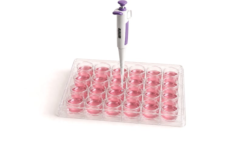
Step 2
Add to culture medium
Add AUMsilence sdASO directly to the culture medium at the study concentration.
Step 3
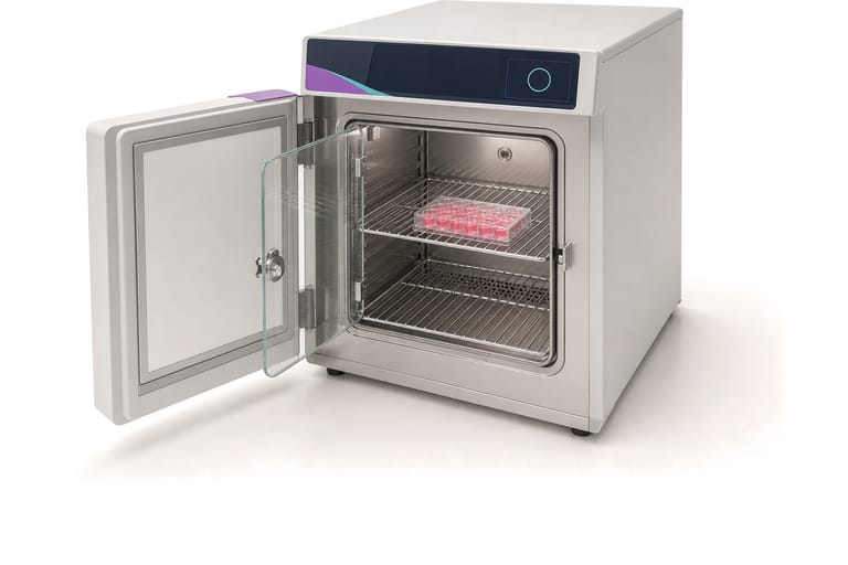
Incubate
Use a model-specific exposure period under standard culture conditions.
- controltreated
Step 4
Quantify knockdown
Quantify target mRNA and, where appropriate, protein abundance or a functional phenotype. Include untreated and non-targeting controls.
Schematic of the three readouts, not data. mRNA by RT-qPCR, protein by immunoblot, protein or phenotype by flow cytometry.


For research use only. Not for use in diagnostic or therapeutic procedures.