

mRNA knockdown
AUMsilence
Self-delivering antisense oligonucleotides for mRNA knockdown, no transfection reagent
AUMsilence
Many primary cells and disease-relevant models are difficult to transfect with conventional methods. Transfection reagents, electroporation, or viral vectors can introduce toxicity, alter the cellular response, or require additional optimization. AUMsilence
- Self-delivery in one step
No transfection reagent, electroporation, or viral vector is required to deliver AUMsilence
sdASOs to cells. - Uptake in difficult-to-transfect cells and in vivo
AUMsilence
sdASOs work in contexts where transfection-dependent methods can struggle. - High specificity and low off-target effects
AUMsilence
sdASOs have high specificity for the intended mRNA. - No cytotoxicity at the optimized working concentration
The working concentration is determined by dose titration for each cell type and target.
Yields and purification
- RPC
- 10 · 25 · 50 · 100 nmol
- HPLC
- 25 · 50 · 100 nmol
- Labeled, HPLC
- 10 · 20 · 25 · 100 nmol · FAM, Cyanine 3, Cyanine 5, Cyanine 7
Prices and lead time are shown on the order page. Custom yields by quote.
At a glance
- Class
- Self-delivering antisense (sdASO)
- Targets
- mRNA knockdown
- Delivery
- Add to culture medium or inject in vivo.
- Fluorescent label
- FAM, Cyanine 3, Cyanine 5 or Cyanine 7
In the lab
The protocol in four steps
The steps below are the AUMsilence
Step 1
Cell preparation
Plate cells in their optimum growth medium at a density appropriate for the cell type. A confluency of 50-70% at the time of treatment is typical.
Step 2
AUMsilence
sdASO stock preparation Prepare AUMsilence
sdASO stock solution by reconstituting lyophilized ASOs at the desired concentration. If you already have a stock solution prepared, skip to Step 3. Step 3
AUMsilence
sdASO delivery to cells Add AUMsilence
sdASO to the cells at the desired final concentration. The recommended working range is 5-20 μM, with a starting concentration of 10 μM. Step 4
Incubation and analysis
Incubate cells with AUMsilence
sdASO and analyze knockdown at appropriate time points. Knockdown is typically observed 24 to 72 hours after treatment, depending on the cell type and the target gene. Longer time points may be required in some systems. Protein knockdown can also be observed from 24 hours after treatment. Where the target protein is long-lived, existing protein must first turn over, so a later time point may be required.
In vitro, the four steps
Step 1
Prepare cells
Seed the cells in a 24-well plate; the oligonucleotide waits in solution.
Step 2
Add to the medium
Add the self-delivering oligonucleotide straight to the medium: no transfection reagent, no carrier.
Step 3
Incubate
Incubate at 37 degrees C in 5% carbon dioxide, humidified, for the exposure time.
Step 4
Quantify knockdown
Measure the knockdown: RT-qPCR, immunoblot, flow cytometry.
Schematic of the three readouts, not data.
Sources
- The pictures are the object pictures of AUM BioTech's workflow slide, rendered views of the objects on a white ground; none is a photograph of an experiment and none records a measurement.
- Tile 04 is a schematic of the three readouts, not data: the two conditions are drawn with a modest difference to show what each readout is, and no magnitude is asserted.
Why
Features of AUMsilence
Self-delivery in one step
No transfection reagent, electroporation, or viral vector is required to deliver AUMsilence
sdASOs to cells. Cells are treated in one step by adding AUMsilence sdASOs to the medium. In primary cells and co-culture systems, where transfection can require optimization, AUMsilence sdASOs need no transfection step. Uptake in difficult-to-transfect cells and in vivo
AUMsilence
sdASOs work in contexts where transfection-dependent methods can struggle. They are taken up by difficult-to-transfect cells and primary cultures with no transfection reagent. AUMsilence sdASOs have been used in T lymphocytes, B cells, neuronal cells and other cells that often take up siRNAs or plasmids poorly. They are also proven in animal models, including mice. High specificity and low off-target effects
AUMsilence
sdASOs have high specificity for the intended mRNA. Cleavage needs the oligonucleotide and the transcript to pair, and a transcript that pairs in part can be bound as well. That is why every candidate sequence is scored for homology across the transcriptome before it is chosen. Unlike siRNA, which can have off-targets via partial "seed" matches through RISC, AUMsilence sdASOs have no RISC-associated off-target effects. No cytotoxicity at the optimized working concentration
The working concentration is determined by dose titration for each cell type and target. At this concentration AUMsilence
sdASOs do not cause cytotoxicity. A reduction in viability reflects the knockdown rather than the chemistry, and the non-targeting control shows whether the knockdown is on target. No transfection reagent, electroporation, or viral vector is required, so the cytotoxicity associated with these delivery methods is eliminated.
Mechanism
Mechanism of action
Antisense binding and RNase H1 cleavage: Each AUMsilence
Nuclear and cytoplasmic targeting: Unlike RNA interference (RNAi) by siRNA, which mainly works in the cytoplasm, AUMsilence
Gymnotic delivery: AUMsilence


Frame 1 of 6: The cell and its medium
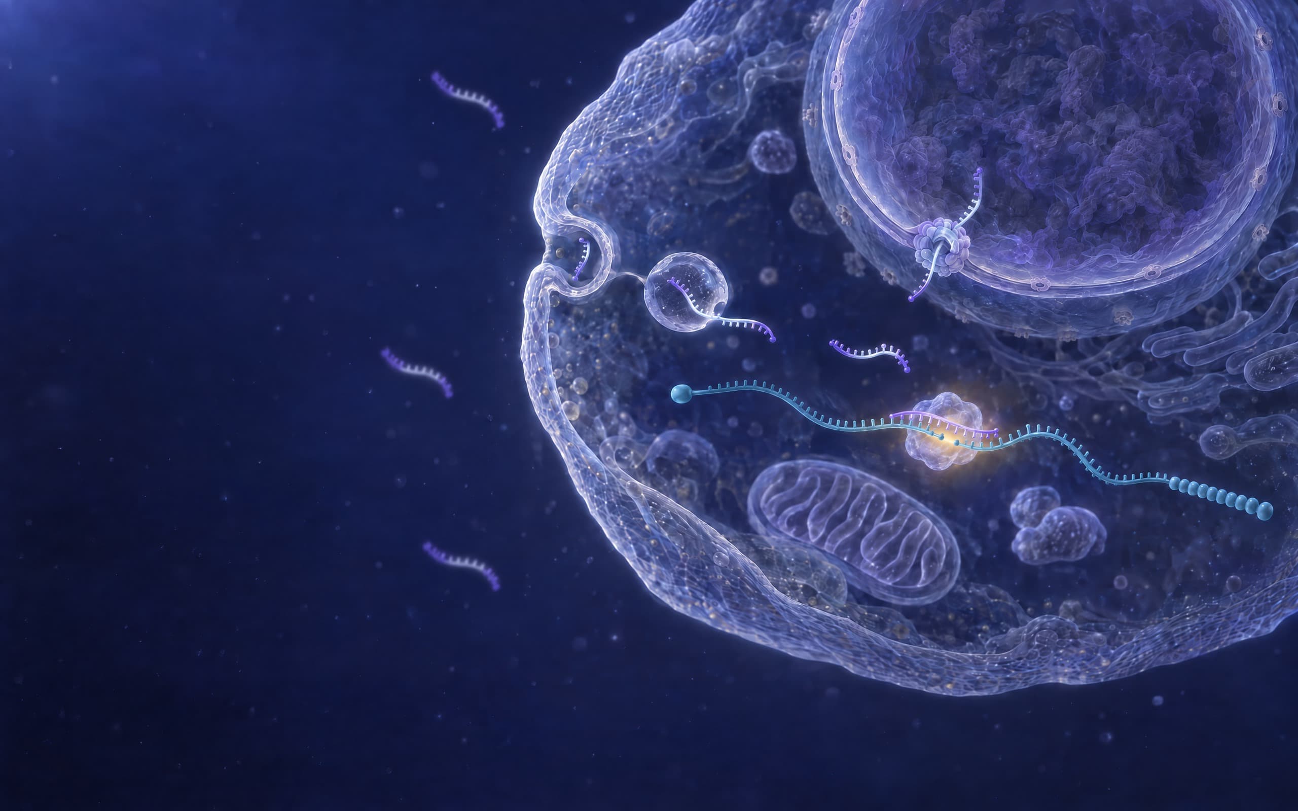
01 The cell and its medium. Self-delivering AUMsilence sdASOs are added to the culture medium as they are, with no transfection reagent, no carrier and no vector; an siRNA duplex or a CRISPR ribonucleoprotein in the same medium needs a reagent, electroporation or a vector to get in. [R01] [R02] [R03] [R04] [R05] [R06] [R07] [R08] [R09]
References
- [R01] Crooke ST, Wang S, Vickers TA, Shen W, Liang XH (2017). Cellular uptake and trafficking of antisense oligonucleotides. Nature Biotechnology 35:230-237. PMID 28244996, doi 10.1038/nbt.3779.
- [R02] Wang S, Allen N, Vickers TA, Revenko AS, Sun H, Liang XH, et al. (2018). Cellular uptake mediated by epidermal growth factor receptor facilitates the intracellular activity of phosphorothioate-modified antisense oligonucleotides. Nucleic Acids Research 46:3579-3594. PMID 29514240, doi 10.1093/nar/gky145.
- [R03] Stein CA, Hansen JB, Lai J, Wu S, Voskresenskiy A, Høg A, et al. (2010). Efficient gene silencing by delivery of locked nucleic acid antisense oligonucleotides, unassisted by transfection reagents. Nucleic Acids Research 38:e3. PMID 19854938, doi 10.1093/nar/gkp841.
- [R04] Dominska M, Dykxhoorn DM (2010). Breaking down the barriers: siRNA delivery and endosome escape. Journal of Cell Science 123:1183-1189. PMID 20356929, doi 10.1242/jcs.066399.
- [R05] Whitehead KA, Langer R, Anderson DG (2009). Knocking down barriers: advances in siRNA delivery. Nature Reviews Drug Discovery 8:129-138. PMID 19180106, doi 10.1038/nrd2742.
- [R06] Falato L, Gestin M, Langel Ü (2021). Cell-Penetrating Peptides Delivering siRNAs: An Overview. Methods in Molecular Biology 2282:329-352. PMID 33928583, doi 10.1007/978-1-0716-1298-9_18.
- [R07] Liang X, Potter J, Kumar S, Zou Y, Quintanilla R, Sridharan M, et al. (2015). Rapid and highly efficient mammalian cell engineering via Cas9 protein transfection. Journal of Biotechnology 208:44-53. PMID 26003884, doi 10.1016/j.jbiotec.2015.04.024.
- [R08] Zuris JA, Thompson DB, Shu Y, Guilinger JP, Bessen JL, Hu JH, et al. (2015). Cationic lipid-mediated delivery of proteins enables efficient protein-based genome editing in vitro and in vivo. Nature Biotechnology 33:73-80. PMID 25357182, doi 10.1038/nbt.3081.
- [R09] Kim S, Kim D, Cho SW, Kim J, Kim JS (2014). Highly efficient RNA-guided genome editing in human cells via delivery of purified Cas9 ribonucleoproteins. Genome Research 24:1012-1019. PMID 24696461, doi 10.1101/gr.171322.113.
- [R10] Beltinger C, Saragovi HU, Smith RM, LeSauteur L, Shah N, DeDionisio L, et al. (1995). Binding, uptake, and intracellular trafficking of phosphorothioate-modified oligodeoxynucleotides. The Journal of Clinical Investigation 95:1814-1823. PMID 7706488, doi 10.1172/jci117860.
- [R11] Juliano RL (2018). Intracellular Trafficking and Endosomal Release of Oligonucleotides: What We Know and What We Don't. Nucleic Acid Therapeutics 28:166-177. PMID 29708838, doi 10.1089/nat.2018.0727.
- [R12] Ochaba J, Powers AF, Tremble KA, Greenlee S, Post NM, Matson JE, et al. (2019). A novel and translational role for autophagy in antisense oligonucleotide trafficking and activity. Nucleic Acids Research 47:11284-11303. PMID 31612951, doi 10.1093/nar/gkz901.
- [R13] Koller E, Vincent TM, Chappell A, De S, Manoharan M, Bennett CF (2011). Mechanisms of single-stranded phosphorothioate modified antisense oligonucleotide accumulation in hepatocytes. Nucleic Acids Research 39:4795-4807. PMID 21345934, doi 10.1093/nar/gkr089.
- [R14] Wang S, Sun H, Tanowitz M, Liang XH, Crooke ST (2017). Intra-endosomal trafficking mediated by lysobisphosphatidic acid contributes to intracellular release of phosphorothioate-modified antisense oligonucleotides. Nucleic Acids Research 45:5309-5322. PMID 28379543, doi 10.1093/nar/gkx231.
- [R15] Cerritelli SM, Crouch RJ (2009). Ribonuclease H: the enzymes in eukaryotes. The FEBS Journal 276:1494-1505. PMID 19228196, doi 10.1111/j.1742-4658.2009.06908.x.
- [R16] Wu H, Lima WF, Zhang H, Fan A, Sun H, Crooke ST (2004). Determination of the role of the human RNase H1 in the pharmacology of DNA-like antisense drugs. The Journal of Biological Chemistry 279:17181-17189. PMID 14960586, doi 10.1074/jbc.m311683200.
- [R17] Liang XH, Sun H, Nichols JG, Crooke ST (2017). RNase H1-Dependent Antisense Oligonucleotides Are Robustly Active in Directing RNA Cleavage in Both the Cytoplasm and the Nucleus. Molecular Therapy 25:2075-2092. PMID 28663102, doi 10.1016/j.ymthe.2017.06.002.
- [R18] Lorenz P, Baker BF, Bennett CF, Spector DL (1998). Phosphorothioate antisense oligonucleotides induce the formation of nuclear bodies. Molecular Biology of the Cell 9:1007-1023. PMID 9571236, doi 10.1091/mbc.9.5.1007.
Applications
Applications and use cases
Gene function studies
Assess the function of a gene by silencing its expression in cells. For example, to study a disease-related gene, treat disease model cells with AUMsilence
sdASO targeting that gene. Changes in phenotype, signaling pathways, or other readouts are then measured. Difficult-to-transfect cells
Use AUMsilence
sdASO to achieve knockdown in primary cells (e.g., primary neurons, hepatocytes, stem cells), suspension cell lines (blood cancer lines, immune cells), or other cells where lipofection efficiency can be low. In vivo gene silencing
AUMsilence
sdASOs are used for in vivo studies with no nanoparticle or viral delivery. They are dissolved in a buffer such as phosphate-buffered saline (PBS) or saline and injected intravenously, intraperitoneally, intrathecally or subcutaneously, depending on your study. High-throughput screening
AUMsilence
sdASOs are used in arrayed screens to identify gene functions or candidate targets. Since no transfection reagent is added, a knockdown experiment scales to 96- or 384-well plate formats.
Every compatible cell type, 3D model, organoid and in vivo route, with the in vitro workflow.
See where it worksExperimental considerations
The working concentration is higher than that of a transfected oligonucleotide. The recommended working range for AUMsilence
In the literature
AUMsilence
Furuhashi et al. · Nature · 2025
Bone marrow niches orchestrate stem-cell hierarchy and immune tolerance
In Nature, a pool of self-delivering AUMsilence sdASOs against Cd200r1 in sorted mouse blood stem cells cut endothelial nitric oxide synthase and abolished long-term reconstitution after transplantation, without changing marrow homing.
Zhang et al. · Science · 2023
TRIM11 protects against tauopathies and is down-regulated in Alzheimer's disease
AUMsilence sdASOs against Trim11, five sequences at 10 μM straight into the medium of primary mouse neurons, entered the cells and lowered TRIM11 to different depths. Seeded tau aggregation worsened by about 50-90%, with about 40% of presynaptic puncta lost.
Schmiedel et al. · Nature Immunology · 2026
Tissue-resident immune cells drive genetic risk in autoimmune and lung diseases
A pool of four AUMsilence sdASOs silenced ZFP57 in primary human macrophages from six donors at 2.0 μM over 10 days. The pool moved 425 transcripts up and 322 down, among them cytokines and chemokines of immune cell trafficking, in a Nature Immunology study of the genetic risk of autoimmune and lung disease.
Marasca et al. · Nature Genetics · 2022
LINE1 are spliced in non-canonical transcript variants to regulate T cell quiescence and exhaustion
AUMsilence sdASOs given by gymnosis at 10 μM, with no transfection reagent, depleted the LINE1-containing transcripts in primary human T cells, restoring the effector function of tumour-infiltrating lymphocytes taken from cancer patients and boosting their killing. They did not restore the proliferation capacity of exhausted T cells.
Chorzalska et al. · Blood · 2018
Bone marrow–specific loss of ABI1 induces myeloproliferative neoplasm with features resembling human myelofibrosis
AUMsilence sdASOs against ABI1, at 15 μM for 48 hours in primary human CD34 positive cells with no transfection reagent, silenced ABI1 by more than 50% and nearly doubled the fraction of cells in S phase.
Mondal et al. · Nature Communications · 2026
Transcription factor 19 modulates fatty acid elongation and unfolded protein response to attenuate palmitic acid-induced hepatic dysfunction
AUMsilence sdASO against Tcf19, injected into the tail vein of mice in two models of fatty liver, cut triglyceride storage and raised inflammation, macrophage invasion and collagen deposition in the liver.
Jin et al. · Science Immunology · 2021
Activation of mTORC1 at late endosomes misdirects T cell fate decision in older individuals
Two independent AUMsilence sdASO sequences against VPS39, added at 1 μM with no transfection reagent, significantly increased the expansion of SARS-CoV-2-reactive and pertussis-reactive human T cells against a matched scramble control.
Xu et al. · Acta Neuropathologica · 2024
MSUT2 regulates tau spreading via adenosinergic signaling mediated ASAP1 pathway in neurons
Self-delivering AUMsilence sdASOs against five messenger RNAs in mouse primary neurons were added straight to the medium at 1 μM. They traced a pathway from MSUT2 through the adenosine A1 receptor to ASAP1 that controls how pathogenic tau seeds get into a neuron.
At the bench
Protocol, controls and what arrives
Add to culture medium or inject in vivo. The protocol, the controls and the How much to order page are written for this product.
- Lead time
- 10-14 business days
- Shipping
- Products are shipped lyophilized, with priority shipping within the United States and international priority shipping elsewhere.
- Quality control
- Every oligo is strictly controlled for quality and is manufactured in an ISO 9001:2015 certified facility.
Order AUMsilence
Provide us with your gene target information, and the AUMsilence platform designs oligonucleotide sequences for your target. Our scientific team is ready to assist with any questions about experimental design or applications.
For research use only. Not for use in diagnostic or therapeutic procedures.