

Protocol
sdASO in vivo protocol
Protocol for self-delivering antisense oligonucleotides in animal models
Self-delivering antisense oligonucleotides (sdASO) are used in animal models.
Protocol overview
The sdASO in vivo application is a five-step process:
- 01Prepare sdASO formulation in appropriate buffer
- 02Select appropriate administration route
- 03Administer to study animals at calculated dosage
- 04Monitor and collect samples at optimal timepoints
- 05Analyze target knockdown in tissues of interest
This protocol can be adapted for different animal models, administration routes, and study endpoints based on your specific research needs.
Key advantages
- No specialized delivery required: Unlike traditional ASOs or siRNAs, sdASO products do not require complex formulations or conjugation for in vivo efficacy
- Multiple administration routes: Tissue uptake has been reported after intravenous, subcutaneous, intraperitoneal and intrathecal injection (the studies are on the publications page)
- Broad tissue distribution: Accumulates in multiple organs and tissues
Administration routes and anatomic sites
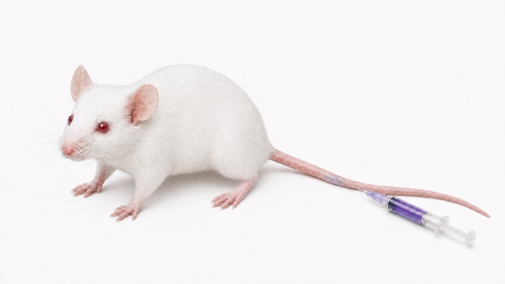
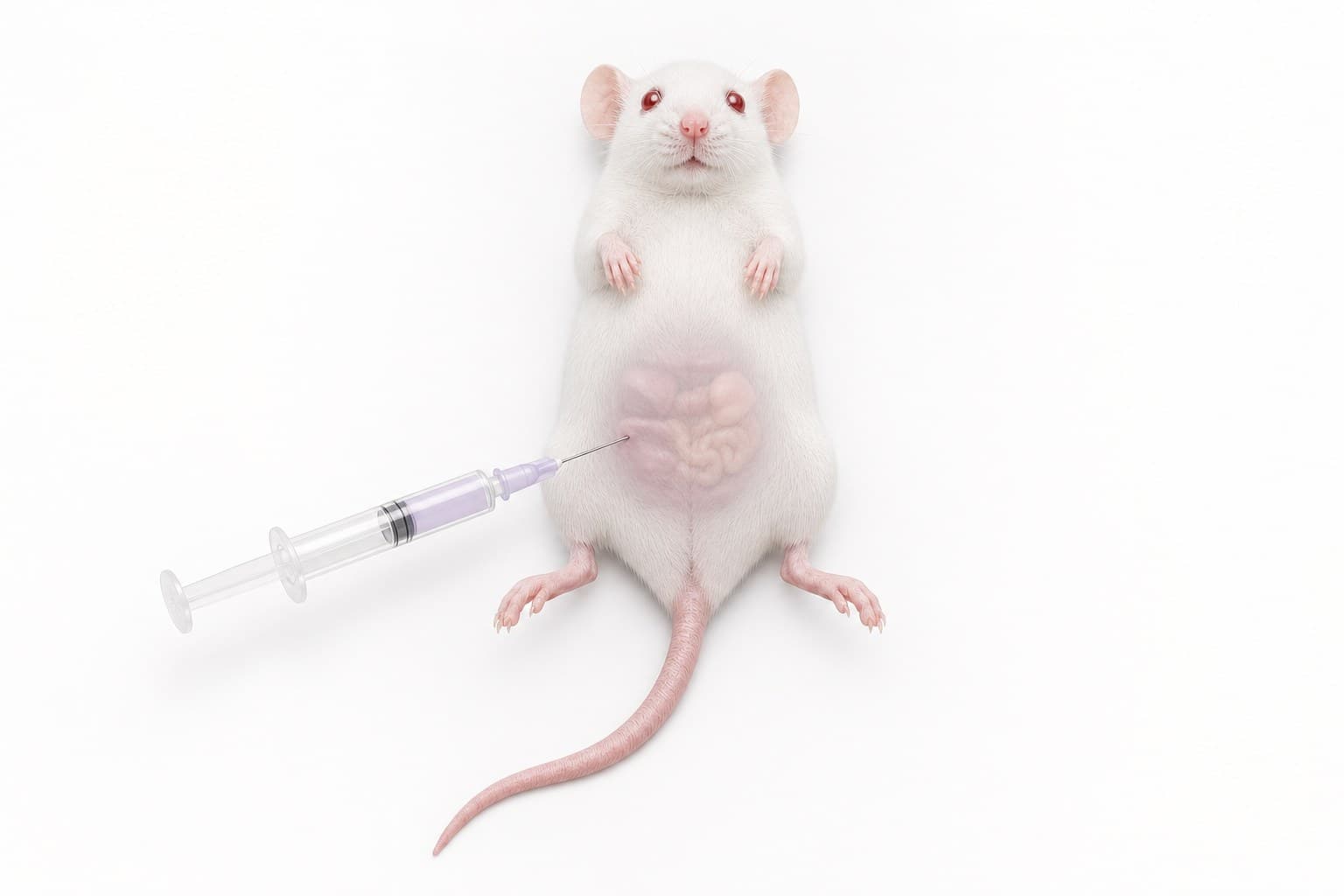
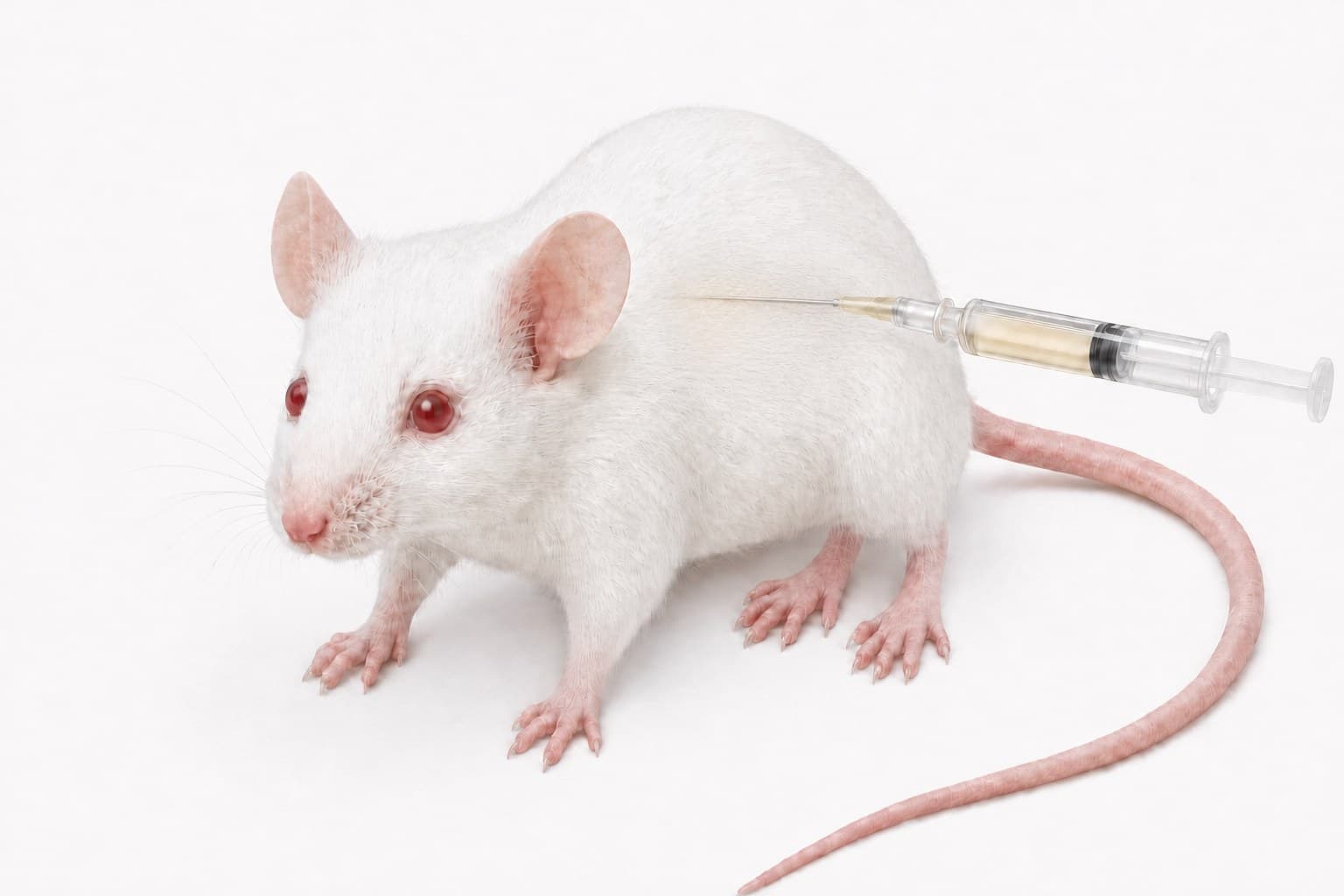
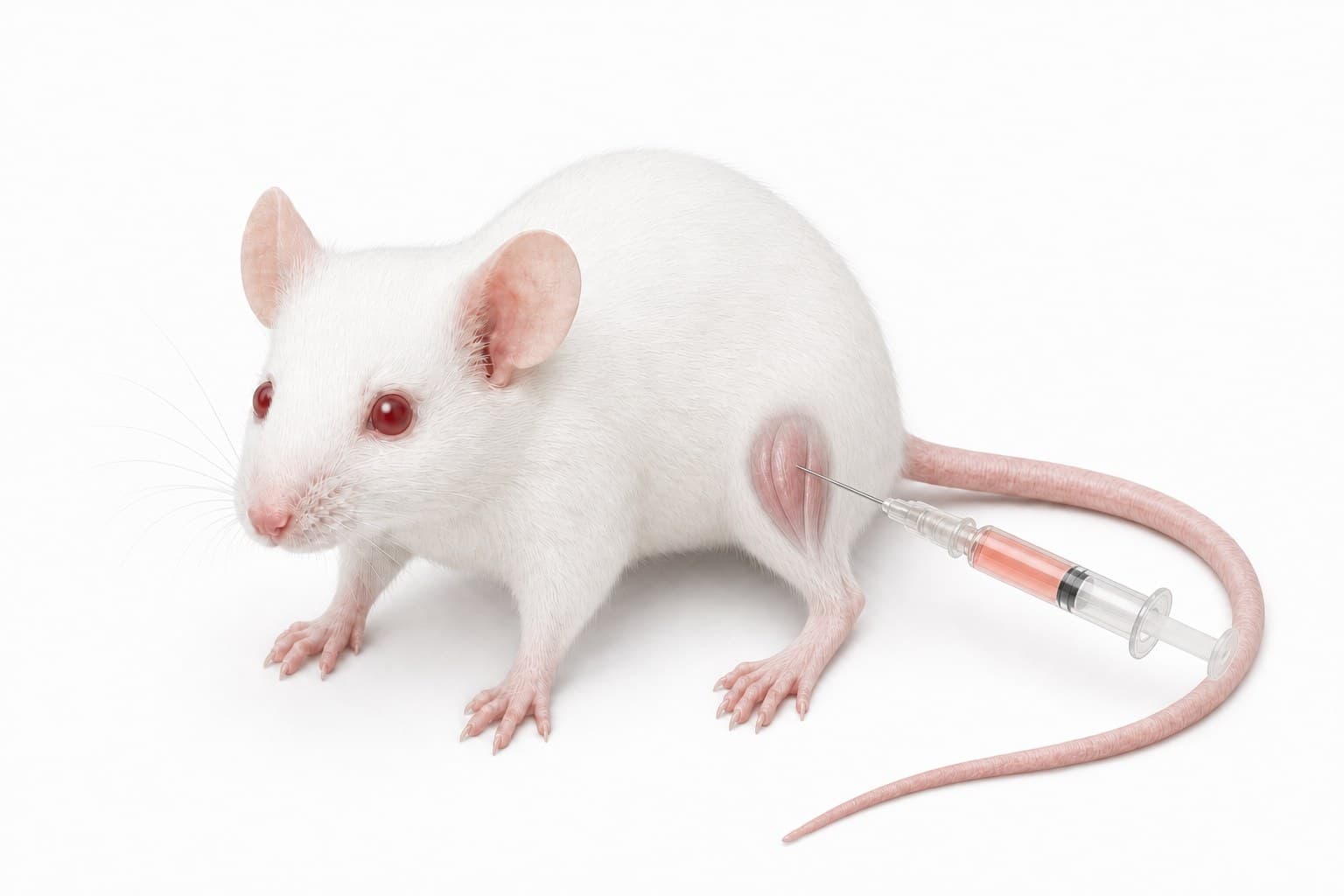
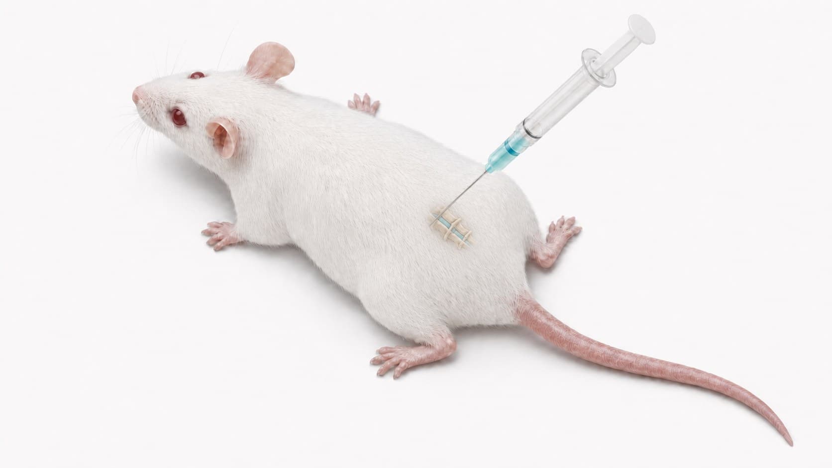
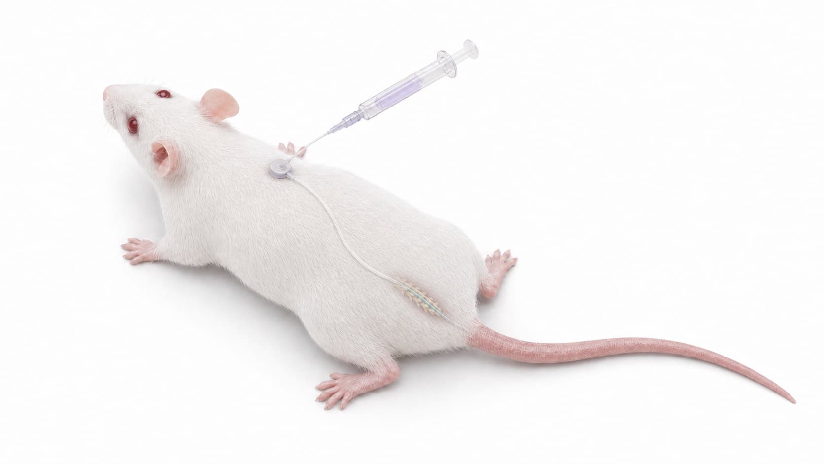
Each route pictured above, with its anatomic site:
- Intravenous, IV: Lateral tail vein. Needle tip positioned within a lateral tail vein and directed proximally.
- Intraperitoneal, IP: Right lower abdominal quadrant. Needle tip placed in the peritoneal cavity through the right lower abdominal quadrant, away from the midline.
- Subcutaneous, SC: Dorsolateral flank. Needle tip positioned beneath intact skin within the subcutaneous tissue plane.
- Intramuscular, IM: Proximal quadriceps muscle. Needle tip positioned in the proximal quadriceps, clear of the posterior sciatic region.
- Intrathecal injection, IT: Lower lumbar L5-L6 interspace. Needle enters through the L5-L6 interspace, immediately cranial to the iliac crest, with the tip in the subarachnoid space.
- Intrathecal catheter, IT: Lumbar catheter and dorsal port. A lumbar intrathecal catheter advances cranially within the subarachnoid space and connects to a dorsal access port.
Materials and reagents
Required items
- AUM BioTech sdASO (lyophilized or stock solution)
- Sterile formulation buffer (PBS, saline, or specialized buffer)
- Administration equipment:
- Sterile syringes and needles (appropriate gauge for selected route)
- Alcohol swabs
- Restrainers (if needed for proper animal handling)
- Animal subjects (appropriate for study design)
- Sample collection materials:
- Surgical tools (for tissue collection at endpoint)
- Blood collection tubes/needles (if performing PK or monitoring)
- RNase-free collection tubes
- RNA preservation reagent (e.g., RNAlater™ or equivalent)
- Liquid nitrogen (for snap freezing samples)
- Analysis materials:
- RNA extraction kit
- qRT-PCR reagents or other RNA analysis tools
- Protein extraction buffers (if analyzing protein knockdown)
- Western blot or ELISA reagents (if applicable)
Detailed protocol
Step 1: Animal preparation
Prepare animals according to institutional guidelines and study requirements.
- Ensure animals have acclimated to facility conditions for at least 5-7 days prior to study initiation.
- Record baseline measurements (weight, behavior, clinical parameters) before administering sdASO.
- Group animals appropriately based on study design (treatment groups, controls, etc.).
- Fast animals if required by your administration protocol (typically not necessary for most routes).
Note: All animal studies should be conducted in accordance with institutional animal care guidelines and with proper ethical approvals. Ensure that personnel are properly trained in animal handling and administration techniques.
Step 2: sdASO formulation preparation
Prepare sdASO formulation at the appropriate concentration for your selected administration route and dosage.
- Resuspend lyophilized sdASO in sterile PBS, saline, or appropriate buffer to the desired concentration.
- For most applications, concentrations of 1-10 mg/mL are suitable for in vivo administration.
- Calculate the required volume based on animal weight and the target dose. AUM's own guidance is to test a dose range, 3-30 mg/kg in mice, to find the dose that silences best. Lower and higher doses may be needed, depending on the administration route and on how long activity has to be sustained. The dosing table below adds a starting dose and a maximum for each animal model.
- Filter sterilize the solution through a 0.22 μm filter if possible.
- Prepare formulations fresh on the day of administration when possible, or store at 4°C for no more than 24 hours.
Important: Avoid multiple freeze-thaw cycles of sdASO formulations. For studies requiring multiple dosing days, prepare fresh formulations each day or store aliquots at -20°C and thaw only once before use.
Step 3: sdASO administration
Administer the sdASO formulation using the appropriate route for your target tissue and research objectives.
- Intravenous (IV) administration: For broader systemic distribution, inject into the tail vein (mice) or appropriate vein according to species. Use a maximum volume of 5-8 mL/kg and inject slowly (over 20-30 seconds).
- Subcutaneous (SC) administration: Inject into the loose skin of the neck or flank. Maximum recommended volume is 10 mL/kg for mice.
- Intraperitoneal (IP) administration: Inject into the peritoneal cavity after proper restraint. Maximum recommended volume is 10 mL/kg for mice.
- Intrathecal (IT) administration: For CNS targeting, inject directly into the intrathecal space. This requires specialized training and typically uses volumes of 5-10 μL in mice.
- Other routes: sdASO can also be administered via local injection to specific tissues (e.g., intramuscular, intraocular, intracerebral) depending on research needs.
Route selection tip: Route selection should be based on your target tissue. IV administration typically yields good distribution to highly perfused tissues (liver, kidney, heart), while SC provides more sustained release. For CNS applications, intrathecal administration puts the oligonucleotide into the cerebrospinal fluid and so bypasses the blood-brain barrier rather than crossing it.
Step 4: Monitoring and sample collection
Monitor animals post-administration and collect samples at appropriate timepoints.
- Monitor animals for any adverse reactions immediately after administration and at regular intervals throughout the study.
- Collect blood samples at predetermined timepoints if pharmacokinetic analysis is part of your study.
- For terminal timepoints, collect target tissues according to your study design. When knockdown is maximal varies with the target, the tissue, the dose and the administration route. Collect at more than one timepoint rather than at a single harvest.
- Process each tissue appropriately:
- For RNA analysis: Either snap freeze in liquid nitrogen or preserve in RNA stabilization reagent
- For protein analysis: Snap freeze or prepare fresh lysates as appropriate
- For histological analysis: Fix in appropriate fixative (e.g., 4% paraformaldehyde)
Note: The kinetics of knockdown vary by tissue and target. Multiple timepoints are recommended for initial studies to establish optimal analysis timepoints.
Step 5: Target knockdown analysis
Analyze collected samples to evaluate the efficacy of sdASO-mediated target knockdown.
- Extract RNA from tissue samples using an appropriate RNA isolation method. Maintain RNase-free conditions to ensure sample quality.
- Perform qRT-PCR to quantify target RNA levels relative to untreated controls and normalized to appropriate housekeeping genes.
- For protein targets, extract proteins and perform Western blot, ELISA, or other appropriate protein quantification methods.
- Analyze functional endpoints relevant to your study (e.g., behavioral assessments, physiological measurements, biochemical parameters).
- If using fluorescently labeled sdASO, tissue distribution can be visualized using fluorescence microscopy on frozen sections.
Analysis tip: It is strongly recommended to include both dose escalation and time course components in pilot studies to determine the optimal dose and timepoint for your specific target and tissue. Knockdown efficacy depends on the target, tissue, dose, and administration route.
Reference guidelines
The tables below provide guidelines for dosing and administration routes:
| Animal model | Typical dose range | Starting dose | Maximum recommended dose |
|---|---|---|---|
| Mouse | 3-30 mg/kg | 5 mg/kg | 50 mg/kg |
| Rat | 2-25 mg/kg | 3 mg/kg | 40 mg/kg |
| Zebrafish (embryo) | 0.1-0.5 nmol/injection | 0.2 nmol/injection | 1 nmol/injection |
| Non-human primate | 1-10 mg/kg | 2 mg/kg | 20 mg/kg |
| Administration route | Target tissues | Advantages | Considerations |
|---|---|---|---|
| Intravenous (IV) | Liver, kidney, spleen, lung, heart | Rapid distribution, high liver uptake | Requires skilled technique, limited CNS distribution |
| Subcutaneous (SC) | Systemic with prolonged exposure | Easy administration, sustained release | Slower onset than IV, may require higher doses |
| Intraperitoneal (IP) | Liver, kidney, intestine, peritoneal tissues | Relatively easy administration | Variable absorption, potential for incorrect administration |
| Intrathecal (IT) | CNS (brain, spinal cord) | Direct CNS access, bypasses BBB | Technically challenging, specialized training required |
| Intramuscular (IM) | Muscle tissue | Local targeting of muscle tissue | Limited distribution beyond injection site |
Dosing notes
- The doses provided are general guidelines; optimal doses may vary depending on the specific sdASO, target, and application.
- For initial studies, a dose-response approach across the guidance range (3-30 mg/kg in mice) is recommended to determine the optimal dose for your target.
- For long-term studies, repeated dosing may be necessary. Typical maintenance regimens include weekly or biweekly administrations at the established effective dose.
Tips and troubleshooting
Optimization tips and best practices
Pilot studies
Multiple controls
Tissue collection
Multi-level analysis
Troubleshooting common issues
Low or variable knockdown efficiency
Adverse reactions or toxicity
Poor tissue distribution
Storage and additional information
Storage conditions
- Store lyophilized sdASO at -20°C upon receipt.
- Reconstituted stock solutions should be stored in single-use aliquots at -20°C.
- Avoid repeated freeze-thaw cycles, as they may affect sdASO stability and efficacy.
- Formulated sdASO solutions for in vivo administration should be prepared fresh when possible. If storage is necessary, keep at 4°C for no more than 24 hours.
Additional considerations
- Duration of effect: Depending on the target and tissue, knockdown typically persists for 1-4 weeks following a single administration.
- Species considerations: While the basic principles apply across species, doses may need adjustment for larger animals.
- Biodistribution: sdASO distribution varies by tissue, with liver, kidney, and spleen typically showing the highest accumulation following systemic administration.
- Pharmacokinetics: sdASO typically have plasma half-lives of several hours to days, with tissue half-lives of days to weeks depending on the chemical modifications.
Note
Request a quote for an in vivo study
A scientist reviews the animal model, the administration route and the dose before an order is placed.
For research use only. Not for use in diagnostic or therapeutic procedures.