

Protocol
AUMsplice
In vitro protocol for self-delivering exon skipping antisense oligonucleotides
AUMskip
Protocol overview
- 01Plate cells at optimal density (50-70% confluency at treatment)
- 02Add AUMsplice
sdASO directly to culture medium - 03Incubate and analyze splice modulation results (typically 24-72 hours after treatment)
This protocol can be adapted for different cell types and various culture vessel formats, from 96-well plates to larger culture vessels.
Key advantages
- No transfection required: Add to media (no lipofection, electroporation, or viral vectors needed)
- Cell compatibility: Works in difficult-to-transfect cell types including primary cells, neurons, and immune cells
- Steric blocking mechanism: Modulates splicing without recruiting RNase H, enabling production of modified proteins
- Low toxicity: Maintains cell viability without transfection reagent-associated toxicity
Materials and reagents
Required items
- AUMsplice
sdASO (lyophilized or stock solution) - Appropriate cell culture medium
- Culture plates or vessels
- Mammalian cells of interest
- Sterile nuclease-free water or buffer (for ASO resuspension)
- Microcentrifuge tubes (for aliquoting ASO stock)
- Standard cell culture equipment:
- Sterile pipettes and tips
- Cell culture hood
- Humidified cell culture incubator
- Centrifuge
- For splice analysis:
- RNA extraction reagents
- RT-PCR reagents
- Primers flanking the target exon
- Gel electrophoresis equipment (for visualization of splice variants)
Detailed protocol
Step 1: Cell preparation
Plate cells in their optimum growth medium at a density appropriate for the cell type. A confluency of 50-70% at the time of treatment is typical.
- For adherent cells: Plate cells the day before treatment, at a seeding density that brings them to that confluency by the time of treatment (or at densities optimized for your specific cell type and assay endpoint). Allow cells to adhere overnight.
- For suspension cells: Prepare cells at appropriate density shortly before treatment with AUMsplice
sdASO. - Optimal cell density will vary with cell type, size, growth characteristics, and the endpoint of your assay.
Step 2: AUMsplice
sdASO stock preparation Prepare AUMsplice
sdASO stock solution by reconstituting lyophilized ASOs at the desired concentration. If you already have a stock solution prepared, skip to Step 3. - Resuspend lyophilized AUMsplice
sdASO using the appropriate volume of sterile nuclease-free water or buffer to achieve the desired stock concentration (typically 1 mM). - Pipette the solution up and down 3-5 times while avoiding the introduction of bubbles.
- Let the vial sit at room temperature for 5-10 minutes to ensure complete resuspension.
- Centrifuge for 30-45 seconds to collect the solution at the bottom of the tube.
- Prepare several aliquots of the stock solution to avoid multiple freeze-thaw cycles.
Important: To avoid degradation, minimize freeze-thaw cycles of your ASO stock. It is strongly recommended to make single-use aliquots of your stock solution and store them at -20°C.
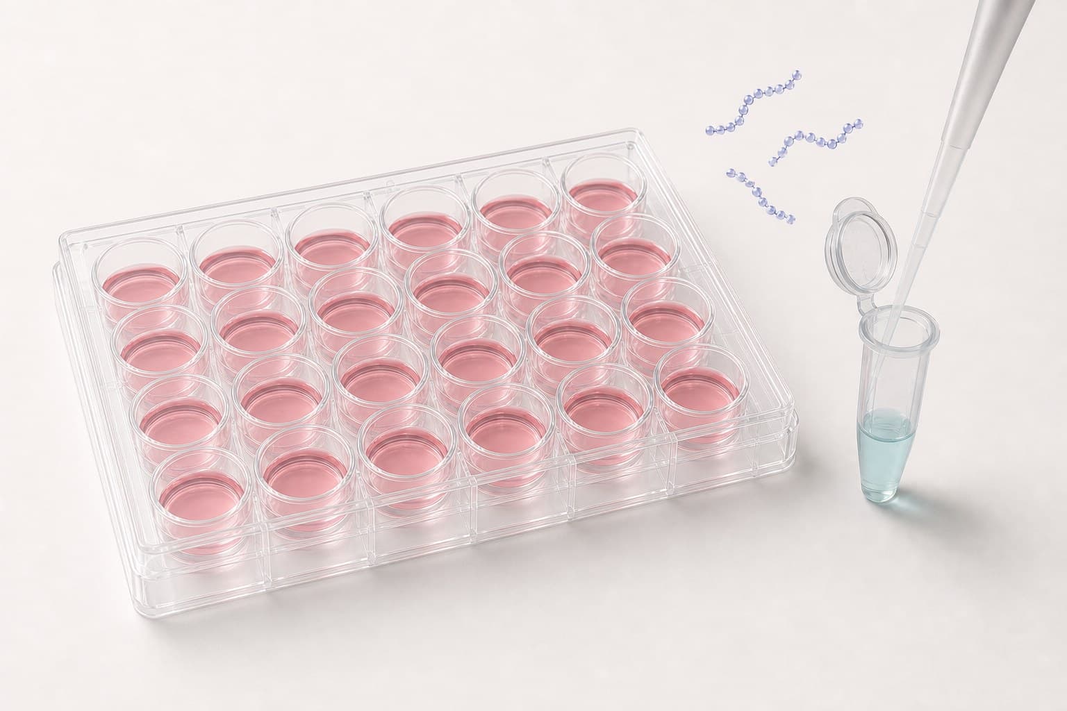
Cells seeded, here in a 24-well plate; the oligonucleotide waits in solution. - Resuspend lyophilized AUMsplice
Step 3: AUMsplice
sdASO delivery to cells Add AUMsplice
sdASO to the cells at the desired final concentration. The recommended working range is 5-20 μM, with a starting concentration of 10 μM. The optimal concentration varies with the target gene, the RNA class (messenger RNA, microRNA or long non-coding RNA) and the cell type, and should be determined by titration for each system. - For adherent cells: Either aspirate the growth media and overlay cells with fresh media containing AUMsplice
sdASO, or add the ASO stock directly to the media overlaying the cells. Mix gently. - For suspension cells: Either pellet the cells by low-speed centrifugation and gently resuspend the cell pellet in media containing AUMsplice
sdASO, or add the ASO stock directly to the media containing the cells. Mix gently. - It is highly recommended to perform a dose optimization using the three concentrations of the range (5 μM, 10 μM and 20 μM) to determine the optimal concentration for your specific exon skipping application.
Optimization tip: An exon may need more than a message does.
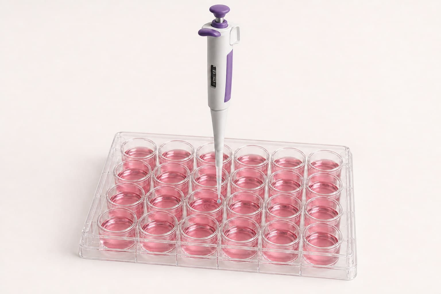
Add the self-delivering oligonucleotide straight to the medium: no transfection reagent. - For adherent cells: Either aspirate the growth media and overlay cells with fresh media containing AUMsplice
Step 4: Incubation and analysis
Incubate cells with AUMsplice
sdASO and analyze splice modulation at appropriate time points. - Return cells to the incubator and maintain under standard culture conditions.
- Analyze AUMsplice
sdASO-treated cells after the desired time point, typically 24-72 hours post-treatment. - RNA splicing analysis can be performed using RT-PCR with primers that flank the target exon, followed by gel electrophoresis to visualize the splice variants (skipped vs. non-skipped).
- For protein-level assessment, Western blot can be performed to detect the altered protein size resulting from exon skipping (typically 48-96 hours post-treatment to allow time for protein synthesis).
Note: For splice analysis, it is critical to design primers that span the skipped exon (e.g., in adjacent exons) to clearly distinguish between splice variants. The exon-skipped product will appear as a faster-migrating band that is smaller by exactly the size of the skipped exon.
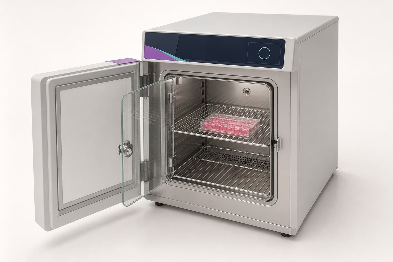
Return the plate to the humidified incubator under standard culture conditions, typically 24-72 hours.
Reference calculations
Making the stock
Reconstitute the vial in nuclease-free water to 1 mM (1 μL per nmol, so 25 μL for a 25 nmol vial). For a 100 μM stock, add 10 μL per nmol.
| Vial | For 1 mM | For 100 μM |
|---|---|---|
| 10 nmol | 10 μL | 100 μL |
| 25 nmol | 25 μL | 250 μL |
| 50 nmol | 50 μL | 500 μL |
| 100 nmol | 100 μL | 1 mL |
Adding the stock to the well
Add the 1 mM stock to the well at 1:200 for 5 μM, 1:100 for 10 μM and 1:50 for 20 μM. At 10 μM that is 1 μL per 100 μL of medium.
| Plate | Medium in the well | 5 μM | 10 μM | 20 μM |
|---|---|---|---|---|
| 96-well | 100 μL | 0.5 μL | 1 μL | 2 μL |
| 48-well | 250 μL | 1.25 μL | 2.5 μL | 5 μL |
| 24-well | 500 μL | 2.5 μL | 5 μL | 10 μL |
| 12-well | 1 mL | 5 μL | 10 μL | 20 μL |
| 6-well | 2 mL | 10 μL | 20 μL | 40 μL |
Treatments per vial
At 10 μM, a 25 nmol vial treats 25 wells of a 96-well plate, 5 of a 24-well plate or 2 of a 12-well plate. Counts are rounded down to whole wells. AUMsplice
| Plate | Medium in the well | 5 μM | 10 μM | 20 μM |
|---|---|---|---|---|
| 96-well | 100 μL | 50 | 25 | 12 |
| 48-well | 250 μL | 20 | 10 | 5 |
| 24-well | 500 μL | 10 | 5 | 2 |
| 12-well | 1 mL | 5 | 2 | 1 |
| 6-well | 2 mL | 2 | 1 | 0 |
Cell density at treatment
Add AUMsplice
Tips and troubleshooting
Optimization tips and best practices
Target site selection
Dose optimization
Extended effect
Controls
Troubleshooting common issues
Low exon skipping efficiency
Difficult detection of splice variants
No protein effect despite RNA skipping
Storage and additional information
Storage conditions
- AUMsplice
sdASOs are shipped in lyophilized form. Upon arrival, store at -20°C. - Resuspended AUMsplice
sdASOs should be stored in aliquots at -20°C to avoid multiple freeze-thaw cycles. - For short-term storage (up to 1 week), resuspended ASOs can be kept at 4°C.
Additional notes
- AUMsplice
sdASOs operate through steric blocking of splice sites rather than RNase H-mediated RNA degradation. - Unlike gene silencing ASOs, AUMsplice
sdASO alters the ratio of splice variants rather than cleaving the target. Total RNA may still fall where the new variant carries a premature termination codon, since the cell degrades such transcripts. Read the variant ratio rather than the total. - No pre-treatment or media change is required before adding AUMsplice
sdASO to cells. - For genetic disease models, confirm that the exon skipping restores the reading frame before proceeding to functional assays.
Note
Request a quote for AUMsplice
A scientist reviews the target exon, the splice site to block and how the variants will be read before an order is placed.
For research use only. Not for use in diagnostic or therapeutic procedures.